Open time: 8 : 00 AM - 5 : 30 PM (Monday - Saturday)
Open time: 8 : 00 AM - 5 : 30 PM (Monday - Saturday)
Available on back-order
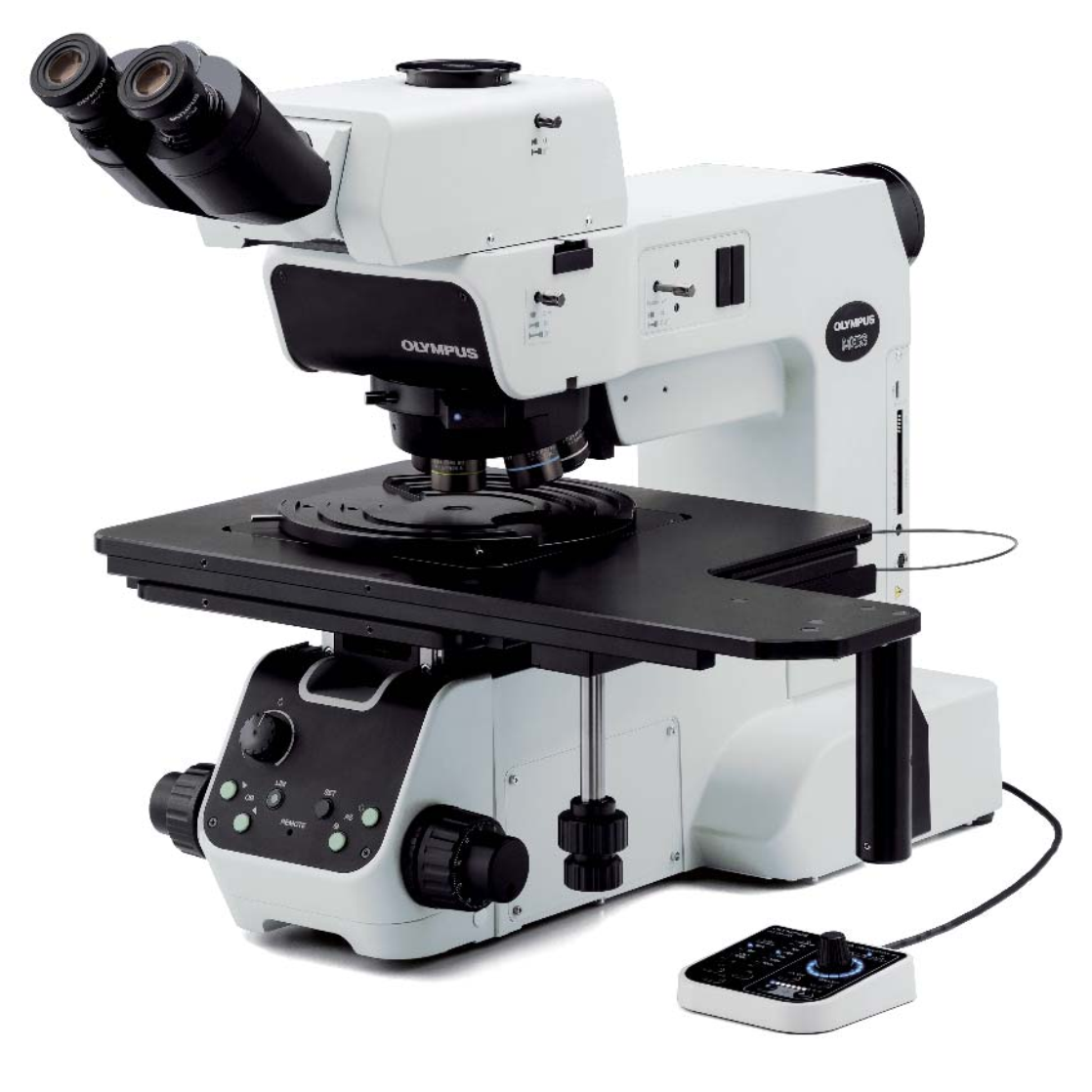
The MX63 series’ various observation capabilities provide clear, sharp images so users can reliably detect defects in their samples. New illumination techniques and image acquisition options within OLYMPUS Stream image analysis software give users more choices for evaluating their samples and documenting their findings.
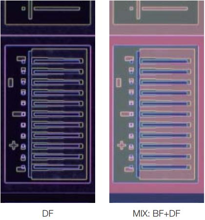
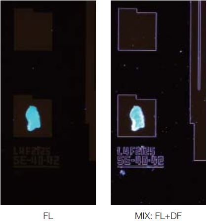
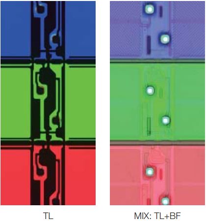
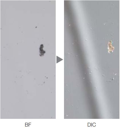
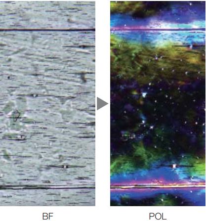
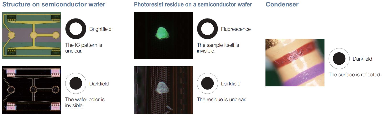
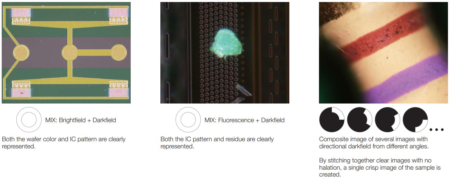
MIX observation technology produces unique observation images by combining darkfield with another observation method, such as brightfield, fluorescence, or polarization. MIX observation enables users to view defects that are difficult to see with conventional microscopes. The circular LED illuminator used for darkfield observation has a directional darkfield function where only one quadrant is illuminated at a given time. This reduces a sample’s halation and is useful for visualizing a sample’s surface texture.

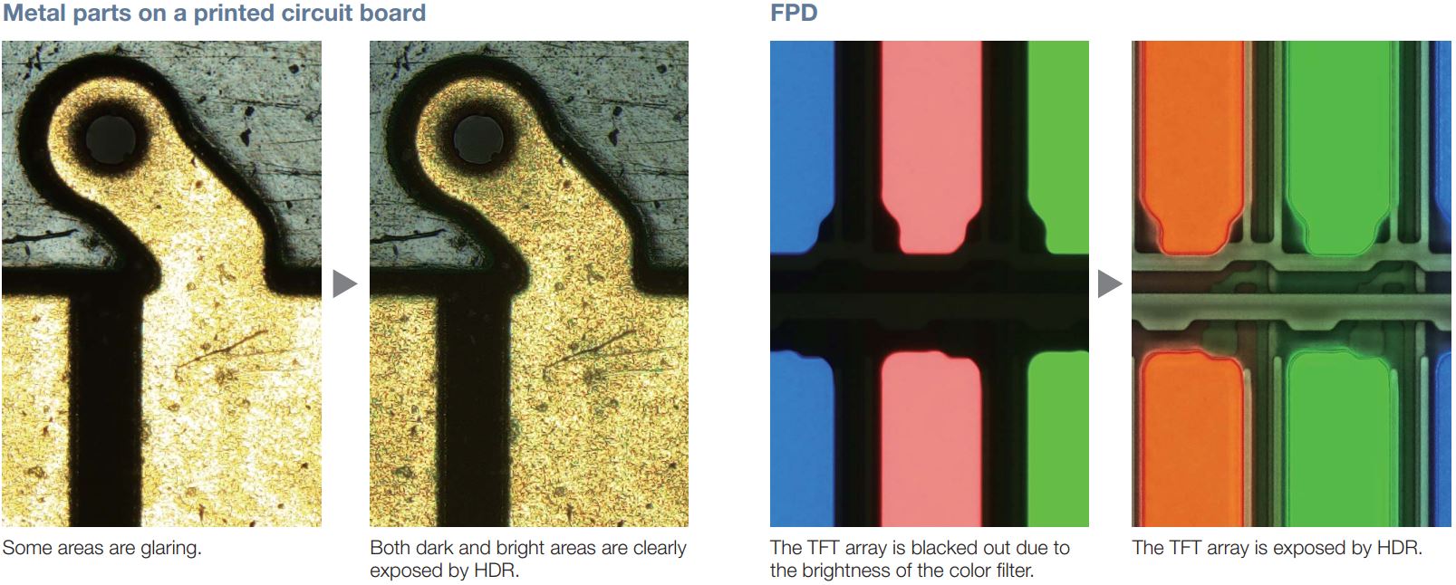
Using advanced image processing, high dynamic range (HDR) adjusts for differences in brightness within an image to reduce glare. HDR improves the visual quality of digital images thereby helping to generate professional-looking reports.
Measurement is essential to quality and process control and inspection. With this in mind, even the entry-level OLYMPUS Stream software package includes a full menu of interactive measurement functions, with all measurement results saved with image files for further documentation. In addition, the OLYMPUS Stream Materials Solution offers an intuitive, workflow-oriented interface for complex image analysis. At the click of a button, image analysis tasks can be executed quickly and precisely. With a considerable reduction in processing time for repeated tasks, operators can concentrate on the inspection at hand.
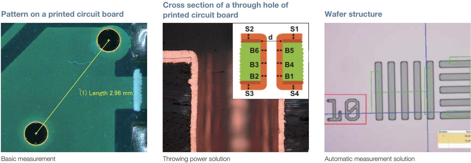
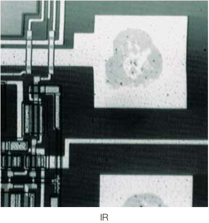
Reflected light microscopy spans a range of applications and industries. These are just a selection of examples of what can be achieved using different observation methods.
|
|
MX63 |
|
|
Optical system |
UIS2 optical system (infinity-corrected system) |
|
|
Microscope frame |
Reflected light illumination |
White LED(with Light Intensity Manager) 12 V 100 W halogen lamp, 100W mercury lamp Brightfield/darkfield/mirror cube manual changeover. (Mirror cube is optional.) 3 position coded mirror units changed by manual operation Built-in motorized aperture diaphragm (Pre-setting for each objective, automatically full open for darkfield) Observation mode: brightfield, darkfield, differential interface contrast (DIC)*1, simple polarizing*1, fluorescence*1, infra-red*1 and MIX observation(4 directional darkfield)*2 *1 Optional mirror cube, *2 MIX observation configuration is required |
|
Transmitted light illumination |
Transmitted light illumination unit: MX-TILLA or MX-TILLB is required. – MX-TILLA: a condenser (NA 0.5) and an aperture stop – MX-TILLB: a condenser (NA 0.6), an aperture stop and a field stop Light source: LG-PS2 (12 V,100 W halogen lamp) Light guide: LG-SF Observation mode: brightfield, simple polarizing |
|
|
Focus |
Stroke: 32 mm Fine stroke per rotation: 100 μm Minimum graduation: 1μm Upper limit stopper and torque adjustment for coarse handle |
|
|
Maximum load weight (including stage and holder) |
8 kg |
|
|
Observation tube |
Wide-field (FN 22 mm) |
Erect and trinocular: U-ETR4 Erect, tilting and trinocular: U-TTR-2 Inverted and trinocular: U-SWTR-3 Inverted and binocular: U-BI30-2 Inverted, tilting and binocular: U-TBI30 |
|
Super-wide-field (FN 26.5 mm) |
Erect, tilting and trinocular: MX-SWETTR (optical path switchover 100% (eyepiece) : 0 (camera) or 0 : 100%) Erect, tilting and trinocular: U-SWETTR (optical path switchover 100% (eyepiece) : 0 (camera) or 20% : 80%) Inverted and trinocular: U-SWTR-3 |
|
|
Motorized nosepiece |
Brightfield Motorized sextuple with a slider slot for DIC: U-D6REMC Motorized centerable quintuple with a slider slot for DIC: U-P5REMC Brightfield and darkfield Motorized sextuple with a slider slot for DIC: U-D6BDREMC Motorized quintuple with a slider slot for DIC: U-D5BDREMC Motorized centerable quintuple with a slider slot for DIC: U-P5BDREMC |
|
|
Stage (X × Y) |
Coaxial right handle with built-in clutch drive: MX-SIC8R Stroke: 210 x 210 mm Transmitted light illumination area: 189 x 189 mm Coaxial right handle with built-in clutch drive: MX-SIC6R2 Stroke: 158 x 158 mm (Reflected light use only) |
|
|
Weight |
Approx. 35.6kg(Microscope frame 26kg) |
|